Finger Tip Injuries
Are very common. The common types of injuries include:
1. crushing injuries eg car door, boot of car, hammer,
2. lacerations eg kitchen knife, circular saw
Tissues Involved:
- Skin
- Finger pulp
- Nerves
- Bone
- Tendons
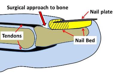
- Nail - See Finger nail injuries
Skin
The skin on the finger tip is very resilient. When skin is lost from injury it needs to be replaced with similar durable skin or the finger tip will continue to split or break down. May times however small skin defects can be allowed to heal with simple dressings and do very well.
Larger defects may require more extensive surgery eg cross finger flaps.
Scars in the finger tips can remain purple and tender for a prolonged period of time.
Finger pulp
The pulp tissue consists of collections of fat surrounded by fibrous bands that create a firm , stable pad for the finger tip to allow grasp and to provide some cushioning to the finger tip
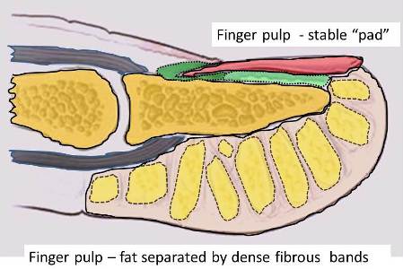
Foreign bodies in the finger pulp can be difficult to locate due to the fibrous bands.
Nerves:
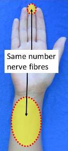
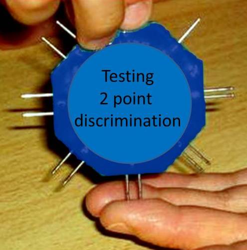
The finger tip is highly sensitive. The nerves are so numerous in the finger pulp that one can distinguish between 2 points only 2mm apart. In the forearm there are far fewer skin nerves. One can distinguish 2 points 15mm apart. See diagram below. There are a similar number of nerve ends in the 2 yellow areas.
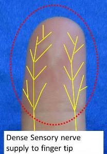
This explains why injuries to the finger tips can be so painful. Lacerations can result in neuroma formation. Hand therapists can teach de-sensitisation exercises to reduce finger tip hypersensitivity. The crushed nerves can make the finger tender for 6 months despite prolonged reduced sensation.
Bone
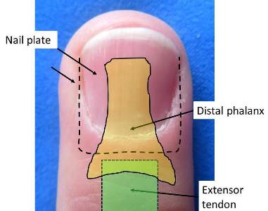
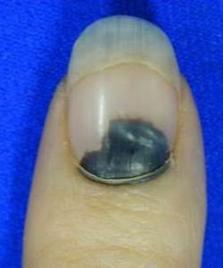
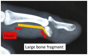
The distal phalanx supports the nailbed. Many fractures are stable because the nail acts as an internal splint. The Periosteum ( Peri = around, Osteum = Bone) is an envelope of tissue around all bones. Crushing injuries often result in multiple fracture lines but the periosteum remains intact. This also increases the fracture stability. Crush injuries can also cause collections of blood under the nail.
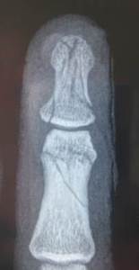
Fractures in the fingers often take 6 weeks to heal but with the distal phalanx it may take 6 – 12 months for the Xray to show healing. More information on finger fractures.
Tendon attachment
Extensor tendons straighten the finger and flexor tendons bend the finger. The extent of the tendon insertion explains the pattern of fractures commonly see in DIP joint injuries.
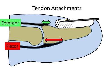

There is only a small gap between the extensor tendon insertion and the base of the nailbed. When operating on mallet finger injuries there is potential to damage the base of the nailbed resulting in nail deformity.